226
E
ELECTROPHORESIS
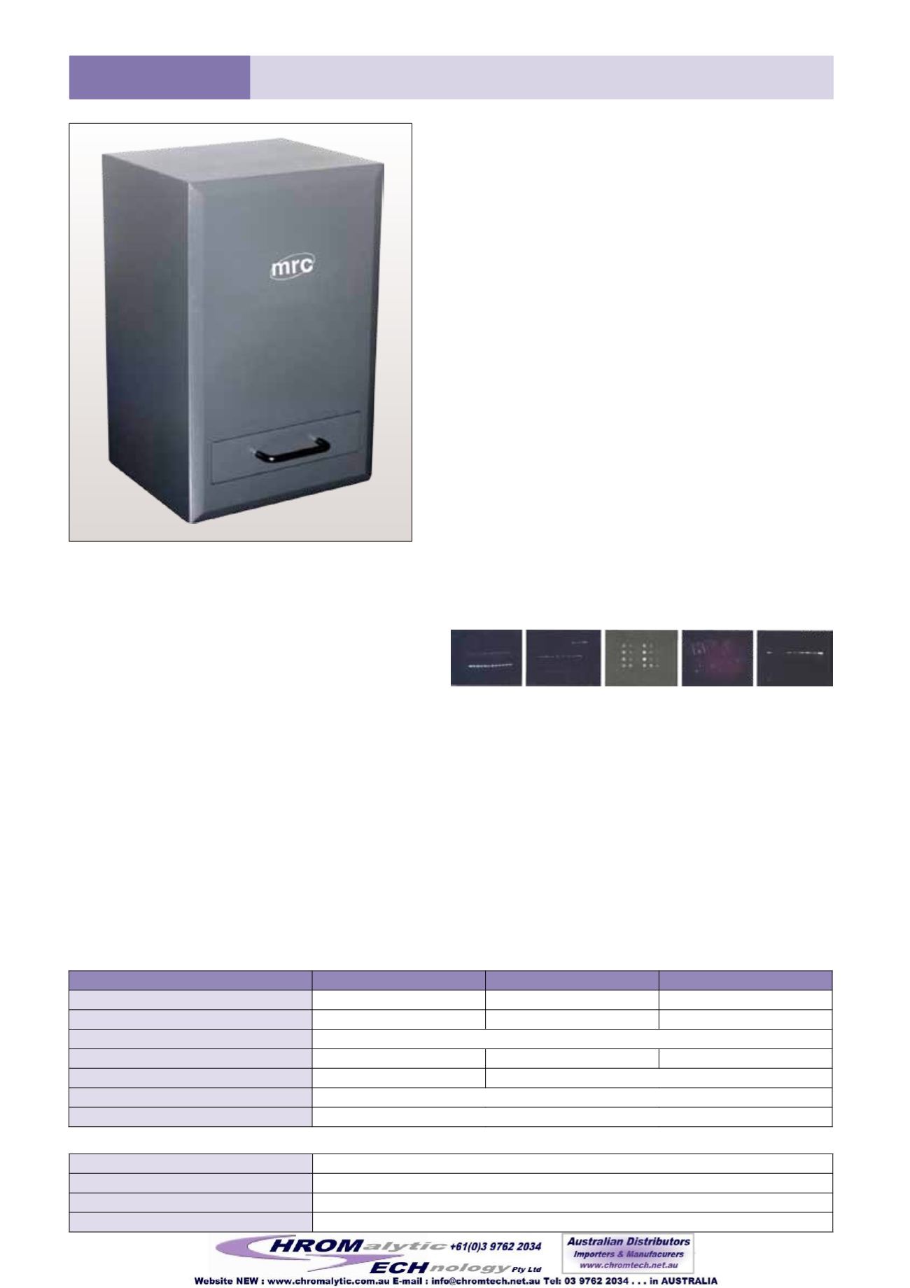
CHS mini Series western blot imaging system is a powerful
scientific grade imaging system that can handle both
Fluorescence and Chemiluminescence imaging
applications. Equipped with a high sensitivity cooled CCD
camera and FO.95 ultra fast lens, the CHS mini offers
the versatility and quality you need in a multipurpose
imaging system with no sacrifice in sensitivity or quality.
The CHS mini offers a complete range of acquisition modes
suitable for imaging the most commonly encountered
fluorescent and chemiluminescent samples.
Features:
●
Three-Stage Peltier Cooled CCD Camera, -60°C bellow
ambient
●
High resolution up to 2048x2048
●
FO.95 Ultra fast lens
●
MUlti-wavelength fluorescent light source
●
Compact frame, convenient operation and light tight
darkroom
●
Easy-to-use capture software and versatile image
analysis software.
Application:
●
Chemiluminescence - Western Lightning, ECL, ECL plus,
CDP Star, Super Signal, CSPD, lumiGlo
●
Nucleic acid detection - Ethidium bromide, SYBR™ Gold,
SYBR™ Green, SYBR™ Safe, GelStar™, Fluorescein, Texas
Red
●
Protein detection - Coomassie blue, Silver Star, Sypro™
Red, Sypro™ Orange, Pro-Q Diamond, Deep Purple™.
CHS-29/32/34 mini, Western Blot Imaging Systems
Model
CHS-29MINI
CHS-32MINI
CHS-34MINI
CCD Size
8.9mm x 6.7mm
11.8mm x 8.9mm
15.2mm x 15.2mm
Pixels
1392x1040, 6.45x6.45um 1600x1200, 7.4x7.4um 2048x2048, 7.4x7.4um
A/D
16bit (65536 grayscale)
CCD Temperature
-55
O
C below ambient
-50
O
C below ambient
-60
O
C below ambient
Lens
17mm F0.95
25mm F0.95
Light Source
EPI-White LED light
Software
Capture & analysis software, free lifetime upgrade
Fluorescent light source
365nm, 395nm, 460nm, 490nm, 530nm, 630nm
Filter Wheel
5 position filter wheel/6 position filter wheel
Filters
530nm, 590nm, 630nm, 670nm
Lens
12mm F1.4, 16mm F1.4, 25mm F1.4, 50mm F1.2
Optional Components
Capture and Anlysis Software:
CHS-Mini Series analysis software is a sophisticated
and intuitive software combining the power of a
comprehensive set of analytical tools and automatic
functions in an easy to use environment.
Analysis Software:
●
Recognize and number gel lanes/bands and
background automatically: add or delete any
band, adjust or move any lane as you want
●
Density contrast: scan appointed gel lane, describe
the scan curve, and then compute the density
integral calculus and peak value of each band in
this lane.
You can also carry on tiny adjustment to light
density measurement scope & contrast several gel
lanes
●
Calculate migration rate of each band,
electrophoretic distances for the molecular weight,
the fragments sizes and the RF values (IEF)
●
Calculate the volume quantification, the height
and the area. You can also compare the volume
of one spot to a reference. Cancel or redo all the
operations infinitely.
Capture Software:
●
Obtain gel image through USB interface/TWAIN interface
●
Live view 16bit original image
●
Time lapse acquisition
●
Control light source and lens through software.
Array Analysis Software:
●
Change the row and col of select area as well as
set dot diameter size
●
Calculate the od value very easily
●
Clear all the data and re-select the test area as
you want
●
Export your analysis results to Excel compatible file
or save your analyzed image on your computer
●
You can preview the report (including company
name and test department, the analyzed image
and data) and print up the results.
Western Blot Imaging Systems


